 Medically Reviewed By: Dr Paul Lim – MBBS, M.Med (Int Med), MRCP (UK), FAMS (Cardiology)
What Does an Abnormal ECG Mean? (Understanding Your Report)
An abnormal ECG means the tracing differs from a textbook pattern — not necessarily that you have heart disease. Concerned about an abnormal ECG reading? Dr Paul Lim will go through your report with you, unhurried, and give you a clear picture of where you stand.
Dr Paul Lim Chun Yih
Senior Consultant Cardiologist & Electrophysiologist
What the Words on Your ECG Report Mean
These are the findings most often printed on a report marked abnormal or borderline. Find the wording on yours and read across. The last column is a general guide to how much each one tends to matter — not an assessment of your result.
| The term on your report | What it means | How much it usually matters |
|---|---|---|
| Right bundle branch block (RBBB) | The signal takes a slightly longer route to the right side of the heart. | Common in otherwise healthy hearts, and more so with age. Usually needs no treatment. |
| Left bundle branch block (LBBB) | The same delay, but on the left side. | Less often harmless than RBBB, so usually worth an echocardiogram to check the heart muscle. New, with chest pain, it is treated as urgent. |
| ST elevation | A stretch of the tracing sits higher than expected. | Can signal a heart attack in progress — an emergency. But it also appears in healthy young people. Your symptoms decide which. |
| ST depression | The same stretch sits lower than expected. | May mean the heart muscle is short of blood, especially with chest pain. Also caused by a fast rate or some medicines. |
| T wave inversion · non-specific T wave abnormality | The wave at the end of each beat points down — an inverted T wave — or does not match a known shape. | Normal in parts of the tracing, and in many younger people. “Non-specific” means the machine could not name it — often nothing. |
| Left or right axis deviation | The overall direction the signal travels through the heart is tilted. | Usually just how you are built. Occasionally points to strain on one side. |
| Sinus tachycardia | A normal heartbeat, running faster than 100 beats a minute. | The rhythm is normal; the question is why it is fast — nerves, fever, dehydration, coffee, low iron, thyroid. See tachycardia. |
| Sinus bradycardia | A normal heartbeat, running slower than 60 beats a minute. | Normal if you are fit, and while you sleep — rates in the 50s are common and usually harmless. It matters more below about 50 in someone who is not an athlete, or if it leaves you tired, dizzy or faint — see bradycardia. |
| Early repolarisation (often printed “early repolarization”) | The tracing lifts early as the heart resets between beats. | A common normal pattern, especially in young, fit adults. Usually harmless, though some shapes are looked at more closely. |
| Atrial or ventricular premature complexes (ectopics) | Extra beats that arrive early, out of step with the rest. | Very common, and usually harmless in an otherwise healthy heart. Worth checking if frequent, or if you feel them as palpitations. |
| First-degree AV block | A slight delay between the top and bottom halves of the heart being triggered. | Rarely causes symptoms and often needs nothing done. Noted so it can be compared later. |
| Left ventricular hypertrophy · left atrial abnormality | A hint that a heart wall is thicker, or a chamber larger, than expected. | An ECG is a poor judge of size and over-calls this in slim people. An echocardiogram gives the real answer. |
These are general descriptions, not an interpretation of your result. The same term can be entirely normal in one person and important in another. A cardiologist reading your full tracing alongside your history and symptoms can tell you what yours means.
What Does “Abnormal” Mean on an ECG Report?
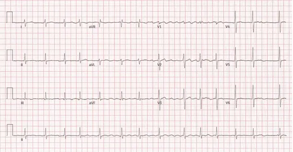
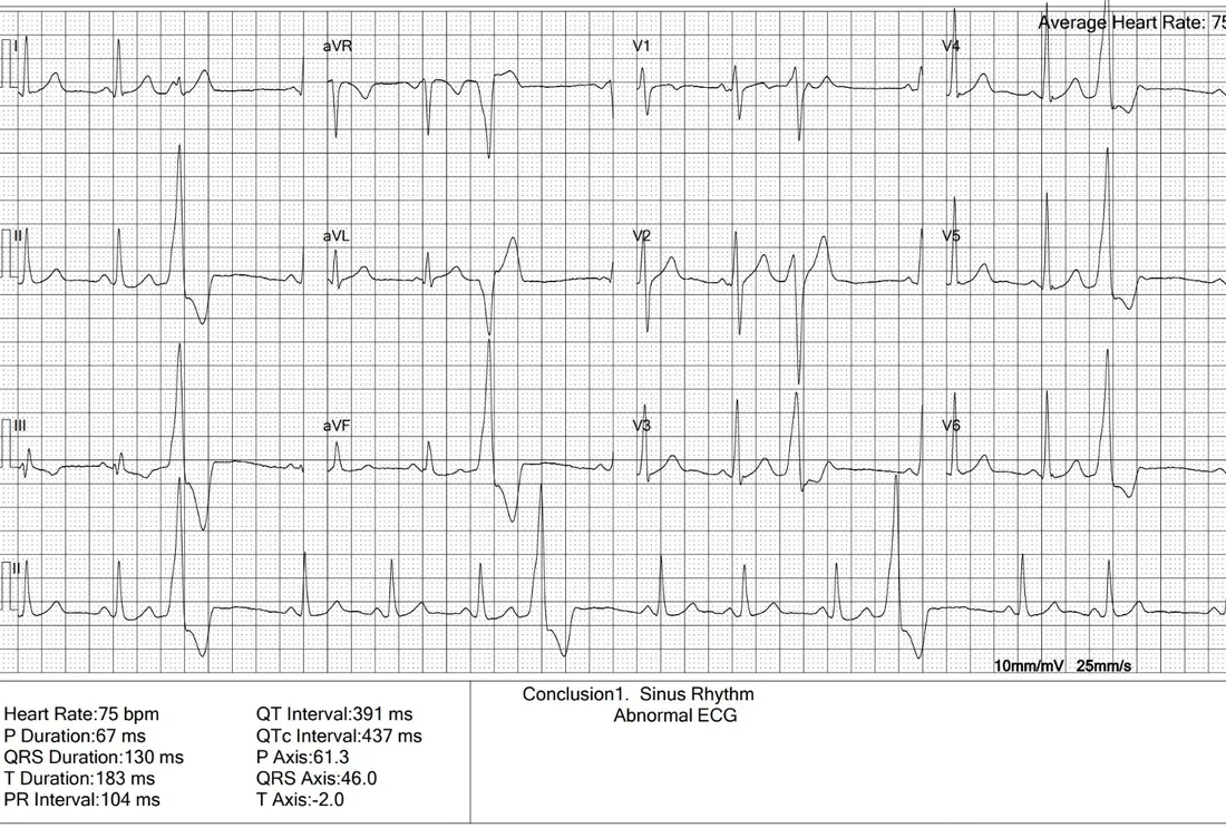
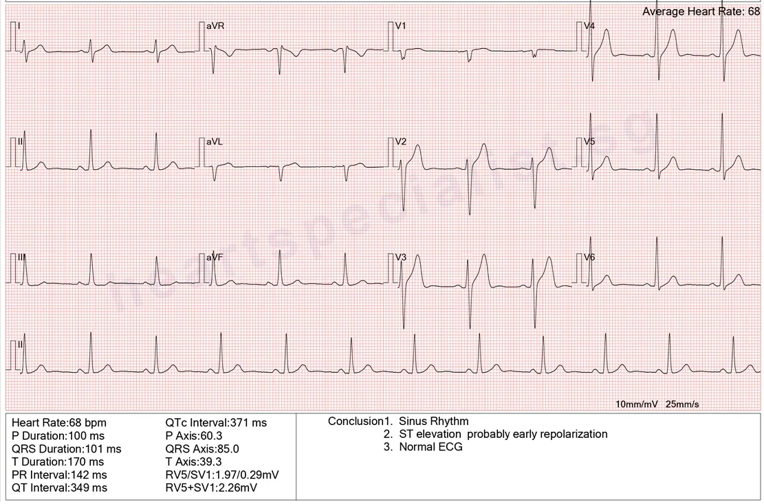
An ECG (electrocardiogram, sometimes written EKG) records your heart’s electrical activity for about ten seconds. A report is marked abnormal when any part of that tracing falls outside the pattern the ECG machine expects — the rate, the rhythm, the shape of the waves, or the timing between them. An abnormal EKG, an abnormal electrocardiogram and an abnormal ECG all mean the same thing; the spellings are just regional.
What Else Can Make an ECG Look Abnormal
A tracing can be flagged for reasons that have nothing to do with heart disease — a reason to interpret it, not to dismiss it.
How it was recorded. Electrodes placed slightly off position are a frequent cause, and moving, talking or shivering adds interference the machine can misread. Repeating the recording resolves a fair number of abnormal results.
Who you are. Build and chest shape change how the signal reaches the skin — which is why slim people are so often flagged for enlargement they do not have. Endurance training remodels and slows the heart, so an athlete’s ECG can look unusual and be perfectly healthy.
What your body is doing that day. Stress, caffeine, nicotine, dehydration, pain, fever or poor sleep all push the rate up, as can an overactive thyroid or anaemia. Shifts in potassium, calcium or magnesium change the waves directly, and several common medicines do too.
Should You Worry About an Abnormal ECG?
What you do next depends far more on your symptoms than on the word printed on the report.
Emergency — Call 995 or go to A&E
Do not wait for an appointment if you have:
- Chest pain, tightness or pressure — especially spreading to the arm, neck or jaw
- Severe breathlessness, especially at rest or lying flat
- Fainting, collapse or near-fainting
- A very fast or irregular heartbeat that will not settle
- Sudden face drooping, arm weakness or slurred speech
Book a cardiologist appointment
See a cardiologist soon if:
- You have palpitations, dizziness, unusual breathlessness or fatigue
- The finding is new — your previous ECGs did not show it
- The report mentions a bundle branch block, ST changes or a rhythm other than sinus
- You have high blood pressure, diabetes or high cholesterol, you smoke, or heart disease runs in the family
- You are concerned about the result, or would like a second opinion
Less likely to be urgent
Worth clarifying, but rarely alarming:
- A borderline label with no specific finding named
- Sinus bradycardia if you exercise regularly and feel well
- A finding unchanged on your ECGs for years
- No symptoms, and no cardiac risk factors
Not sure which applies to you? Speak to a cardiologist rather than guess from the report.
Concerned about your ECG? Have it reviewed by a cardiologist.
How an Abnormal ECG Is Sorted Out
Most are resolved in one visit. Where they are not, the next tests are straightforward.
-
1
The tracing is read properly
The full 12-lead tracing is read against your symptoms, medicines and family history — not the machine’s summary. Bring any earlier ECGs: a finding unchanged for years reads very differently from a new one.
-
2
The ECG is often repeated
A fresh recording with careful electrode placement, taken while you are settled, clears a good number of abnormal readings on the spot.
-
3
Further tests only where they add something
If the rhythm is the concern, a Holter monitor catches what ten seconds cannot. If the structure is, a 2D echocardiogram shows it directly — and the two tests answer quite different questions. If blood supply is, a treadmill stress test or CT coronary angiogram answers it better.
-
4
You leave knowing where you stand
Whether the finding turns out to be normal for you, worth watching, or worth treating, you leave knowing which — with a written summary if you need one.

Clinical Experience
Reviewed by Dr Paul Lim
Senior Consultant Cardiologist & Cardiac Electrophysiologist
Dr Paul Lim is a Senior Consultant Cardiologist who subspecialises in the heart’s electrical system — the part of cardiology an ECG actually measures. Reading tracings and working out which findings matter is core to that work. He completed advanced fellowship training at Barts Heart Centre, London under Singapore’s HMDP award, and regularly sees patients who want an abnormal ECG properly explained, or a second opinion on one.
Have Your ECG Report Reviewed
Bring your tracing to a consultation with Dr Paul Lim, whether you are concerned about it or would simply like a second opinion.
Send Us a Message

Where to Get an Abnormal ECG Reviewed in Singapore

Opening Hours
| Monday – Friday | 8:30 AM – 5:30 PM |
| Saturday | 8:30 AM – 12:30 PM |
| Sunday & Public Holidays | Closed |
By Appointment Only
Abnormal ECG: FAQ
The questions patients ask most after being handed a report marked abnormal.
Does an abnormal ECG mean I have heart disease?
Usually not. A report is marked abnormal whenever the tracing differs from a textbook pattern, and many healthy people produce tracings that do exactly that. Some findings do matter, particularly alongside chest pain, breathlessness, palpitations or fainting. The word tells you the tracing was not textbook — not that something is wrong.
Can stress or anxiety cause an abnormal ECG?
Yes, commonly. They raise the heart rate enough to be reported as sinus tachycardia — a normal rhythm running fast, not a rhythm disorder. Caffeine, nicotine, dehydration, fever, pain, poor sleep, an overactive thyroid and anaemia do the same. Anxiety is a genuine explanation, but not one to assume before the others have been considered.
What does a borderline ECG mean?
It means a measurement sat just outside the machine’s normal range without being clearly outside it. Borderline describes where a number fell — not a diagnosis, and not a milder version of abnormal. Many borderline tracings prove entirely normal for that person on review.
Can an ECG be wrong?
The recording is accurate; the interpretation printed on top often is not. It comes from the machine matching your tracing against stored patterns, with no knowledge of your age, build, symptoms or previous ECGs. Electrodes slightly out of position or muscle movement distort a trace too — which is why a repeat sometimes looks entirely different.
Will an ECG show a blocked artery?
Not reliably. It shows a heart attack happening now or one that has already occurred, but a narrowed artery not currently limiting blood flow often produces a normal tracing. A normal ECG does not rule out coronary artery disease — a treadmill stress test, CT calcium score or CT coronary angiogram answers that far better.
My ECG was abnormal but my stress test was normal — what does that mean?
A common and generally reassuring combination: the resting tracing has a feature that is simply normal for you, and your heart behaved well under exertion. It does not close the matter entirely, because a stress test is not designed to assess a resting finding such as a conduction delay. Your cardiologist reads the pair together.
Do I need to repeat the ECG?
Often, yes — it is one of the most useful next steps. A repeat with correct electrode placement resolves a good number of abnormal readings on its own, and comparing it against an older tracing is more informative still. Bring any previous ECGs to your appointment.
Where This Guidance Comes From
This page is educational and is not a diagnosis. A tracing is interpreted alongside your symptoms, history and previous ECGs — which is what a cardiology consultation is for.